Biological age is linked to age-related brain changes
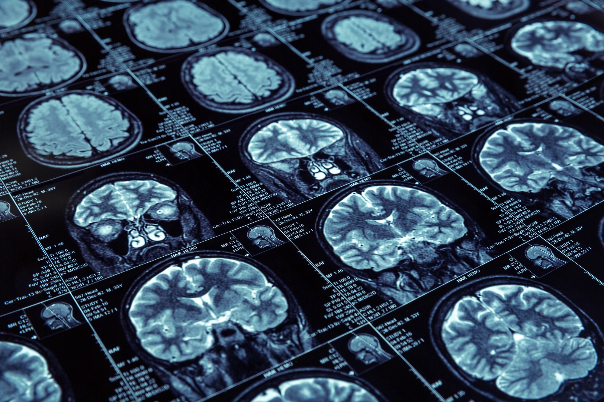
It is a universally accepted truth that our body changes as we age. As people go over a certain age, their bones become more brittle, they develop wrinkles, and they get sick more often. Our brain changes as well. Though these changes may not be as noticeable, they can be detected with multiple methods available to scientists. Specialists can perform multiple tests from evaluating our reflexes to measuring brain waves and MRI scans. The latter approach is a favorite of researchers because it can give insight into the brain function and detailed brain structure.
Brain Aging
One of the problems with evaluating how the brain changes with age is the fact that the brain is a very complex organ. It has multiple parts, and each part is responsible for a certain number of functions. We usually imagine the brain as this big, complex organ consisting of two hemispheres adorned with deep grooves, like a labyrinth. In fact, this image of the brain is actually only one of its parts – the cerebral cortex. It is also one of the most interesting ones because the cerebral cortex is acting like a supercomputer, getting enormous input from all over the body, processing it and sending out multiple orders. The cerebral cortex has two major layers – the so-called grey matter and white matter. Grey matter is the “working” area. It consists of nerve cells or neurons that accept and send out signals. Neurons in their turn have long branches that connect to other cells and other brain areas, making multiple connections and forming the white matter, the “communications” area. Both structures are crucial for normal brain activity and are influenced by the process of aging.
One of the hallmarks of aging process is the increase in the number of cells that die. Cells die in the brain, too. As a result, the grey matter of the cortex-the cortical layer – becomes progressively thinner. MRI scans have shown that as people advance in age, their cortical layer slowly loses density. It can also happen when a person is suffering from dementia or other types of neurodegenerative disorder.
What is interesting is that if you take two elderly people, of a similar age and health, their cortices may not be changed to a similar degree. In one person, the loss of cells would be significantly lower than in another.
One of the explanations for such an occurrence is that there is a difference between chronological age (i.e, number of years a person has lived) and biological age (the actual level of aging of the body). There are multiple ways to evaluate biological age. One of the newest and most reliable is evaluating the level of DNA methylation.
Our DNA is not just a double helix. It’s four nucleotides that form the genetic code can have other chemical moieties attached. These additions can have a significant impact on the activity of certain genes. DNA methylation is such a mark; one of the 4 nucleotides called cytosine has a methyl (CH3) group attached to it. DNA methylation plays multiple roles in the functioning of the genome. Recently, it was also shown that the level of DNA methylation in the cells of the body is very closely linked with age (1). Based on this finding, a new method of evaluation of biological age – so-called DNAm clocks, or epigenetic clock-was developed. To calculate the biological age with this method, the researcher needs to see how many so-called CpG positions in DNA in a certain number of genes have a methyl group attached to them.
It was already known that biological age increases when a person has developed some significant health problems, such as cancer (2) or heart disease (3). Until recently, while the link between biological age and the state of the brain in adult healthy people was unknown.
A team of researchers from the University Of Nebraska Medical Center in Omaha (4) has decided to change that. To achieve this goal, they have decided to examine the possibility that biological age and cortical thickness in the brain are connected. To do that, they tested 82 adults, all healthy, aged 22-72. Each person had two main tests performed: a blood test to look at the methylation of their DNA and an MRI scan to investigate the condition of the subject’s brain. Then the researchers compared several measures:
The difference between the chronological and biological ages of each personThe level of cortical thickness in each patientThe relationship between the biological age and cortical thickness in each patient
At the end of the study, the team had observed several interesting phenomena. First, biological age of women was slightly lower than the biological age of the men. It implies that women are slightly younger biologically than men, though the difference was not considered statistically significant. Second, they have also established that the people who were older biologically had a thinner cerebral cortex. Moreover, certain areas were affected more than others, for example:
The frontal region of the cortex (5) that is responsible for decision making and so-called “executive functions”. This region is basically responsible for delegating and coordinating complex tasks across different brain areas. Superior temporal region (6) responsible for processing sounds Inferior parietal cortex (7) – this area is responsible for understanding emotions, gathering information about the senses and also for facial recognitionMedial occipital cortex (8) – it helps us understand the depth of objects and evaluate distances. This area also contributes to the development of memory.
Besides those major areas, the regions responsible for controlling movement and detecting internal signals from the body also had thinner cortex layer.
With an increase in age, the thinning has spread to additional areas besides the ones mentioned above:
Bilateral superior temporal cortex (9) that is responsible for processing soundsOther areas are responsible for movement control and evaluation of signals from the body.
Age Acceleration
Another important finding was made in a group of people whose biological age was significantly higher than the chronological one. Their internal clocks were running faster. This condition is called age acceleration. In subjects with age acceleration, the thinning of the cortex was even more widespread and it has affected the following areas:
Left orbitofrontal cortex – the function of this area is not particularly clear and is supposed to be involved in decision makingPosterior superior temporal gyrus – responsible for producing and understanding language, evaluating sounds, and also assisting in memory formation Left parahippocampal gyrus (10) – contributes to creating memoriesFusiform gyrus (11) – an important structure that participates in memory and also understanding visual informationRight temporal sulcus (12) – a structure involved in day-to-day social actions.
Other areas responsible for the senses were also affected by cortical thinning in this group.
Though this study used only two parameters to evaluate the changes related to age in the brain, it has several very important implications. First, it is another report that corroborates the utility of epigenetic clock for evaluating age. It is evident that only chronological age would not be a reliable risk factor for age-related disease. DNAm clock, on the other hand, reflects the actual condition of the individual and the aging process they undergo more accurately. Another important aspect of this study is that it provides an explanation as to why is there is so much variability in brain function in people of the same age.
Why is it important to measure biological age accurately? Mainly because aging is associated with certain disorders, and measures of the epigenetic clock can help determine if this person is at risk of developing them. As was already mentioned earlier, biological age is accelerated in people with cancer, cardiovascular diseases, as well as neurodegenerative diseases such as Alzheimer’s, Parkinson’s, and Huntington’s disorders. And in the latter cases, cortical thinning is often observed, because one of the main symptoms of these disorders is the loss of the neurons, especially in certain areas. As it was reliably proven that higher biological age implies a decrease in the thickness of the cortex, it is also plausible to see this parameter as a sign that conditions associated with neural deterioration are also likely to present or are likely to develop.
Age acceleration described in the present study can also be used as such a marker. As the results of MRI scans suggest, the cortical thinning is often located in the areas responsible for decisions, recognition, movement, memory, sight and speech. And usually, we observe problems with the same functions in the elderly: difficulties with coordination, movements, making decisions, controlling emotions, or recognizing family members. Those problems could obviously be more exacerbated in people with age acceleration. And as rule, age acceleration must have an underlying cause – dementia disorder, infection, or possible cancer. Therefore, if the doctor knows that the patient’s biological age is significantly higher than normal, he or she could reliably assume that the person is at risk of having neuron loss and probably some serious issue affecting his health and normal functioning of their body.
This study is the first of its kind to look at the relationship between cortical thickness and DNAm clock values. Still, previously there was a study (13) that looked at another brain structure – white matter. In this study, the researchers have established that the structure of white matter was changed in people with epigenetic age acceleration. Taken together, these data suggest that aging involves disruptions in multiple brain structures, and application of the DNAm clock can be used as a biological marker that points to the possibility that a number of such changes are either already present or will likely soon develop.
Despite these useful results, the authors admit that their study has limitations. As their test subjects were living people, they could not just take samples from their brains and see if the DNA methylation levels were like the ones observed in the blood. Still, previous studies found that the values of DNAm clocks in various tissues of the body were similar, so it is possible that the analysis of the brain tissue, in this case, may not have brought any additional information.
This study is hopefully the first in a line of new studies that would help us understand how the brain is affected during the aging process and whether the changes in the brain could be reversible. There is some evidence (14) that it is possible to reverse epigenetic changes – by understanding the link between methylation, biological age, and brain deterioration, scientists may find new clues and avenues to treat brain disorders.
References
Horvath, S. DNA methylation age of human tissues and cell types. Genome Biol 14, 3156 (2013). https://doi.org/10.1186/gb-2013-14-10-r115DNA methylome analysis identifies accelerated epigenetic ageing associated with postmenopausal breast cancer susceptibility. Ambatipudi, Srikant et al. European Journal of Cancer, Volume 75, 299 – 307 https://doi.org/10.1016/j.ejca.2017.01.014Perna, L., Zhang, Y., Mons, U. et al. Epigenetic age acceleration predicts cancer, cardiovascular, and all-cause mortality in a German case cohort. Clin Epigenet 8, 64 (2016). https://doi.org/10.1186/s13148-016-0228-zProskovec AL, Rezich MT, O’Neill J, et al. Association of Epigenetic Metrics of Biological Age With Cortical Thickness. JAMA Netw Open. 2020;3(9):e2015428. doi:10.1001/jamanetworkopen.2020.15428https://doi.org/10.1016/B978-008045046-9.00960-8Karnath, H. New insights into the functions of the superior temporal cortex. Nat Rev Neurosci 2, 568–576 (2001). https://doi.org/10.1038/35086057Yoshie, M., Nagai, Y., Critchley, H. et al. Why I tense up when you watch me: Inferior parietal cortex mediates an audience’s influence on motor performance. Sci Rep 6, 19305 (2016). https://doi.org/10.1038/srep19305Rehman A, Al Khalili Y. Neuroanatomy, Occipital Lobe. [Updated 2020 Jul 31]. In: StatPearls [Internet]. Treasure Island (FL): StatPearls Publishing; 2020 Jan-. Available from: https://www.ncbi.nlm.nih.gov/books/NBK544320/Karnath, H. New insights into the functions of the superior temporal cortex. Nat Rev Neurosci 2, 568–576 (2001). https://doi.org/10.1038/35086057https://doi.org/10.3389/fnhum.2015.00431https://dx.doi.org/10.1016%2Fj.neuropsychologia.2015.06.033Shultz S, Lee SM, Pelphrey K, McCarthy G. The posterior superior temporal sulcus is sensitive to the outcome of human and non-human goal-directed actions. Soc Cogn Affect Neurosci. 2011;6(5):602-611. doi:10.1093/scan/nsq087Epigenetic Age Acceleration Assessed with Human White-Matter Images. Karen Hodgson, Melanie A. Carless, Hemant Kulkarni, Joanne E. Curran, Emma Sprooten, Emma E. Knowles, Samuel Mathias, Harald H.H. Göring, Nailin Yao, Rene L. Olvera, Peter T. Fox, Laura Almasy, Ravi Duggirala, John Blangero and David C. Glahn. Journal of Neuroscience 3 May 2017, 37 (18) 4735-4743; DOI: https://doi.org/10.1523/JNEUROSCI.0177-17.2017Horvath, S., Raj, K. DNA methylation-based biomarkers and the epigenetic clock theory of ageing. Nat Rev Genet 19, 371–384 (2018). https://doi.org/10.1038/s41576-018-0004-3
Recent Blog Posts
-
 05 Apr 2024Addressing Prostate Cancer Surge: EpiMedTech Global's Response
 -
 23 Mar 2024The Transformative Journey of Pregnancy: A Deeper Look into Maternal Biological Aging
 -
 11 Mar 2024Unlocking the Connection Between the Gut Microbiome, Muscle Function, and Cognition in the Elderly: Insights from the PROMOTe Trial
 -
 02 Mar 2024Turning Back Time with Your Fork: The Fasting-Mimicking Diet Unveiled
 -
 14 Feb 2024Understanding the Secrets of Bat Longevity: A Glimpse into DNA Methylation
 -
 29 Dec 2023Redefining Alzheimer's Management: How Time-Restricted Feeding Aligns Body Clocks and Enhances Brain Health
 -
 26 Dec 2023Unlocking the Mystery of Cervical Cancer: The Vital Role of Folate
 -
 26 Nov 2023Unlocking the Secrets of Age Reversal: How Cutting Edge Research is Paving the Way